
Multi Radiance has spent over 20 years focused on research, clinical studies, and developing technologies specific for your patients and your practice. MyVetLaser.com is an organized library of all that information and more
Request Access Today! →


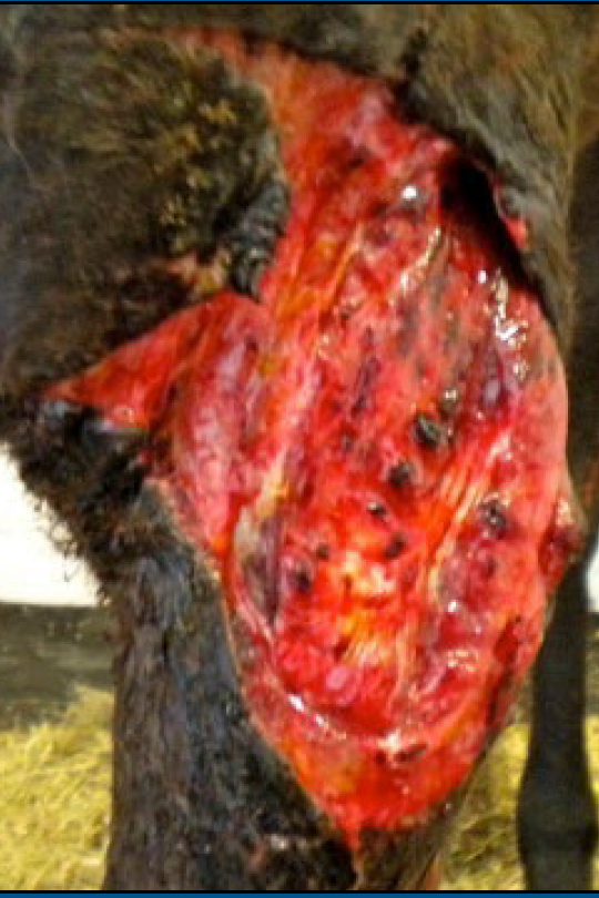
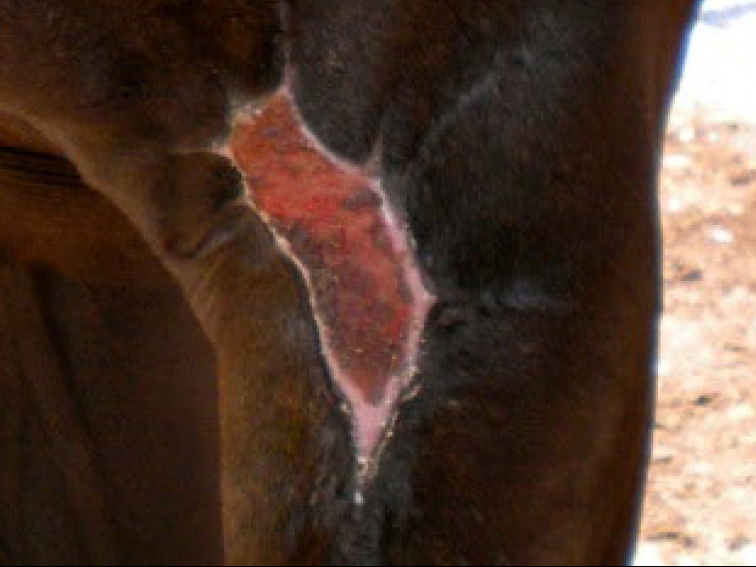
Presentation:
- Lacerations, abrasions, contusions, penetrating, contaminated, and chronic nonhealing wounds.
- Post-op incisions are included in this section because they are a “surgical wound.” “Incisional wounds.”
Mechanism of Action:
Wound healing is a natural multi-staged process in which the patient responds to any traumatic incident with an orchestrated biochemical cascade of events set in motion to repair the damage and restore these disrupted tissues to normal anatomical activity while leaving little cosmetic evidence of their initial occurrence.
This repair and restoration event has four overlapping phases:
- Hemostasis (Clotting)
- Inflammation
- Tissue growth (Proliferation)
- Maturation
Laser therapy significantly influences each of these stages, accelerating each phase of the healing process. The biochemical cascade of events resulting from the application of laser therapy has the ability to cause:
- Vasoconstriction
- Activate fibroblasts and macrophages.
- Release growth factors and neurotransmitter substances.
- Influence collagen synthesis and organization.
- Increase vascularization.
- Aid in the proliferation of epithelial cells.
- Initiate tissue remodeling.
Treatment:
- Cleaned, debridement, and lavage.
- Pain management
- Systemic antibiotics
- Closure of the wound with sutures, staples, glue, or surgery.
- Laser therapy is administered immediately after closure followed by appropriate sequential therapy sessions.
Open Wound:
- Administer laser therapy immediately after cleaning, debridement, and lavage before applying topicals, moist dressings, or bandages.
- Administer at each bandage change after cleaning and before the application of topicals and dressings.
- Topicals
- Bandaging
- Controlled exercise.
Protocol Instructions
Priority Principle: Wound Repair
- Initiate treatment after cessation of hemorrhage
- Painful or contaminated: treat off-contact
- Tolerant: treat on contact with emphasis on margins
- Apply Blue Light therapy after cleaning and debriding.
- Saturate entire area for 3-5 minutes
- Apply 1000-3000 Hz, off contact, scanning technique, 1 cm/sec.
- 80% of the therapy session should be applied around the margins of the wound
For More Specific/Custom Wound Applications
General Guidelines: Daily treatment for consecutive days, up to three times per day, until pain and swelling are resolved, then increase the interval between therapy sessions.
- Blue light: 100%, scanning technique, 4 -5 minutes over entire wound, up to 3X/day.
- Initial swelling: 1000-3000 Hz, scanning technique, entire wound surface and well into wound margins.
- Pain management: sessions of 1000 Hz, then 1000-3000 Hz, then 5000 Hz as needed.
- When swelling and pain are receding, apply tissue repair, PROM exercises, and functional stress protocols to accelerate healing.
- Scanning over the epithelial margins will encourage cellular migration.
- 1-250 Hz every other day or every three days.`Once the wound has granulated, to avoid more granulation tissue, avoid scanning over the wound bed.
| Abrasions/Contusions | ||
| Symptoms | Protocol | Blue Light |
| Inflammation | 50 Hz | 1-2 Minutes | 100% 1/2" Above with Dome Probe and Scanning | 5 Minutes |
| Pain | 1000 Hz | As Needed | 100% 1/2" Above with Dome Probe and Scanning | 5 Minutes |
| Tissue Repair | 1-250 Hz | 3-5 Minutes | 100% 1/2" Above with Dome Probe and Scanning | 5 Minutes |
*Each case is unique and will require appropriate protocol adjustments.
| Puncture (Not Infected) | ||
| Symptoms | Protocol | Blue Light |
| Inflammation | 50 Hz | 1-3 Minutes scanning over affected areas |
Application upon discretion |
| Pain |
1000 Hz | 1 Minutes 1000-3000 Hz | 2 Minutes 5000 Hz | 1 Minute |
Application upon discretion |
| Tissue Repair | 1-250 Hz | Over entire area | Application upon discretion |
| Puncture (Infected) | ||
| Symptoms | Protocol | Blue Light |
| Inflammation | 50 Hz | 1-3 Minutes scanning over affected areas |
Application after cleaning, Scanning entire area |
| Pain |
1000 Hz | 4 Minutes 1000-3000 Hz | 3 Minutes 5000 Hz | 1 Minute |
Application after cleaning, Scanning entire area |
| Tissue Repair | 1-250 Hz | Over entire area | Application after cleaning, Scanning entire area |
*Each case is unique and will require appropriate protocol adjustments.
OPEN
| Symptoms | Protocol | Blue Light |
| Inflammation | 50 Hz | 1-3 Minutes scanning over affected Areas |
Scanning applications for 5 minutes; up to 3X per day |
| Pain |
1000 Hz | 4 Minutes 1000-3000 Hz | 3 Minutes 5000 Hz | 1 Minute |
Scanning applications for 5 minutes; up to 3X per day |
| Tissue Repair | 1-250 Hz | 2-3 Minutes | Scanning applications for 5 minutes; up to 3X per day |
SUTURED
| Symptoms | Protocol | Blue Light |
| Inflammation | 50 Hz | 1-3 Minutes scanning over affected Areas |
Scanning applications for 5 minutes; up to 3X per day |
| Pain |
1000 Hz | 4 Minutes 1000-3000 Hz | 3 Minutes 5000 Hz | 1 Minute |
Scanning applications for 5 minutes; up to 3X per day |
| Tissue Repair | 1-250 Hz | 2-3 Minutes | Scanning applications for 5 minutes; up to 3X per day |
*Each case is unique and will require appropriate protocol adjustments.


